Ultrasound uses sound waves to create safe, radiation-free images — speak with your doctor about the right scan for your symptoms.
An ultrasound scan is a medical test that uses high-frequency sound waves to capture live images from the inside of your body. It's also known as sonography.
The technology is similar to that used by sonar and radar, which help the military detect planes and ships. An ultrasound allows your doctor to see problems with organs, vessels, and tissues without needing to make an incision.
Unlike other imaging techniques, ultrasound uses no radiation. For this reason, it's the preferred method for viewing a developing fetus during pregnancy.
Pelvic Ultrasound
This type of ultrasound is considered a diagnostic tool. It can examine the uterus, bladder, or prostate. A pelvic ultrasound can diagnose abnormal bleeding, pain, menstrual problems, ovarian cysts, fibroids, kidney or bladder stones, and cancer in the uterus or ovaries.
Abdominal Ultrasounds
This type of ultrasound is useful in examining organs like the liver, gallbladder, spleen, pancreas, bile ducts, kidneys, and bladder. It aids in the diagnosis of many conditions or to assess damage caused by illnesses. It can also help to determine abdominal pain, stones in the gallbladder, identify the cause of an enlarged organ, blood clots, or plaque buildup.
Obstetric Ultrasounds
Used routinely during pregnancy to monitor the growth of the fetus, this is one type of ultrasound that many people are familiar with, though you may also know it as a fetal ultrasound. With this test, physicians can learn the age of the fetus and diagnose any possible abnormalities, and, in addition, determine if there's more than one fetus.
Transvaginal Ultrasound
This diagnostic ultrasound requires that a woman empty her bladder before it can be performed. The transducer is inserted just a few inches inside the vagina, and it is commonly used when a patient has unexplained pelvic pain.
Transrectal Ultrasound
This type of ultrasound is used to observe the prostate gland. The transducer is inserted into the rectum in order to have the sound waves travel to the prostate. The transducer is moved around in order to observe images from several different angles.
Carotid and Abdominal Aorta Ultrasound
This is used to monitor the carotid artery in the neck that carries blood from the neck to the brain. It is primarily used to screen patients for stenosis, which is a blockage or narrowing of the carotid artery. A stenosis can greatly increase the patient's risk of a stroke. A carotid and abdominal aorta ultrasound will also identify if an aneurysm is present, which is an abnormal enlargement of the aorta.
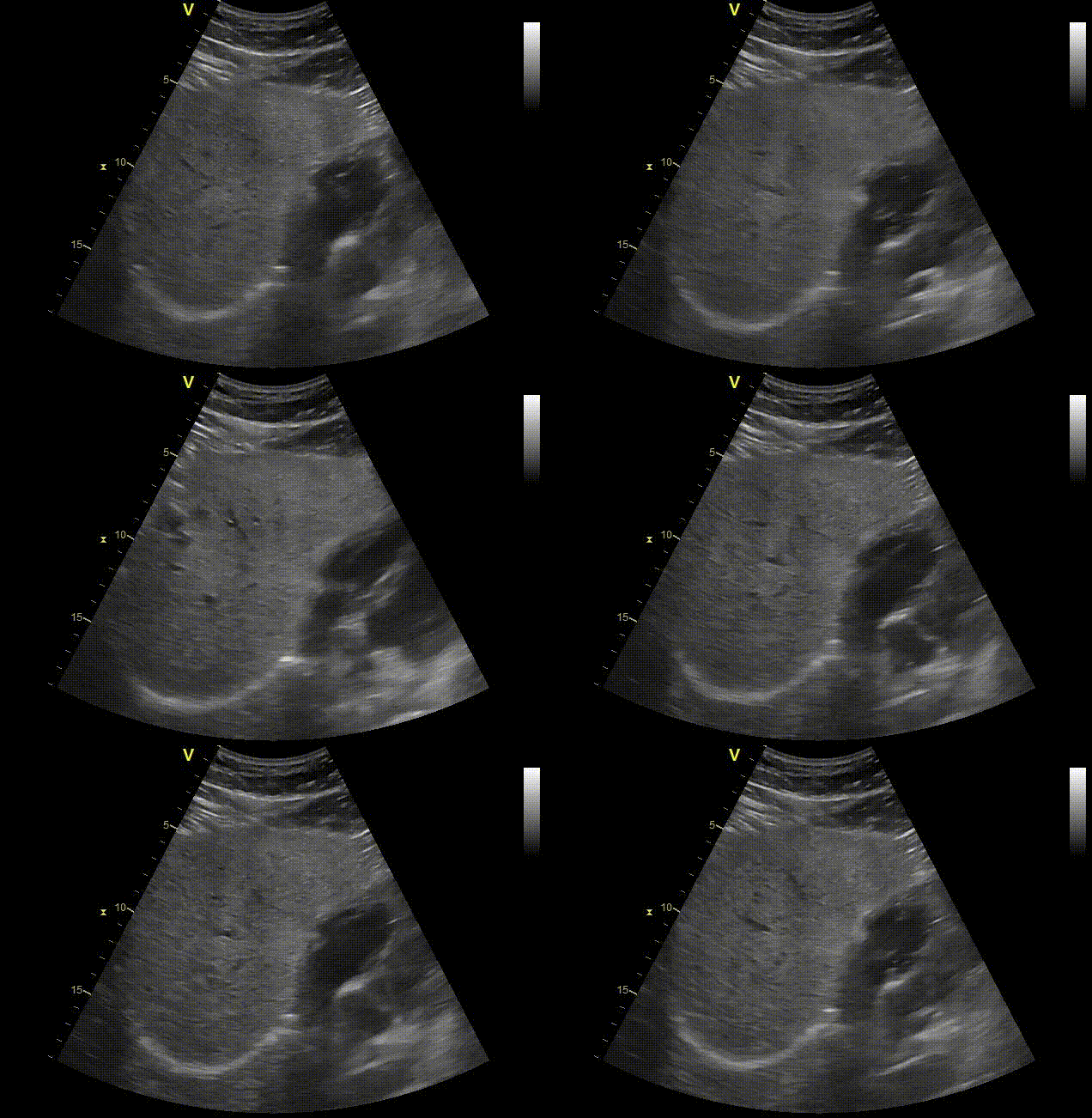
Abdominal ultrasound of the liver may show cysts, masses, blocked blood vessels, and dilated bile ducts. Cysts are confidently diagnosed with ultrasound. Many masses need further testing with CT or MRI for diagnosis. Dilated bile ducts are well seen with ultrasound. We often do not know the cause based on ultrasound alone. Additional testing with MRI may be needed.
Abdominal ultrasound of the gallbladder is one of the best tests for the gallbladder. We can see sludge, stones, masses and inflammation of the gallbladder or cholecystitis. Gallstones are a common cause of pain in the right upper abdomen. Cholecystitis is when the gallbladder becomes inflamed.
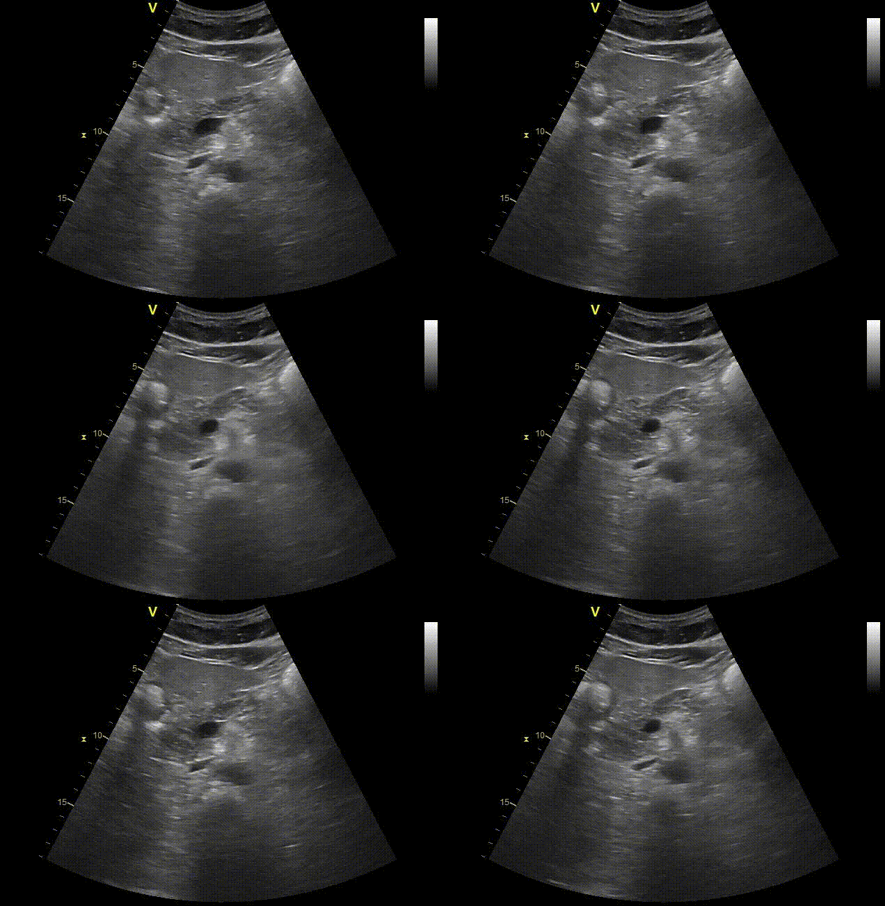
Abdominal ultrasound of the pancreas can show us cysts, masses, a dilated pancreatic duct, collections around the pancreas to name some. The pancreas is sometimes not well seen because it is a deep structure and located behind bowel loops.
We can see the spleen which is in the left side of the abdomen. The spleen helps with fighting infections in the blood and filters the blood. The spleen can have cysts, masses, infarcts, and infections. Ultrasound can sometimes identify abnormalities but further testing may be needed to say what the finding is.
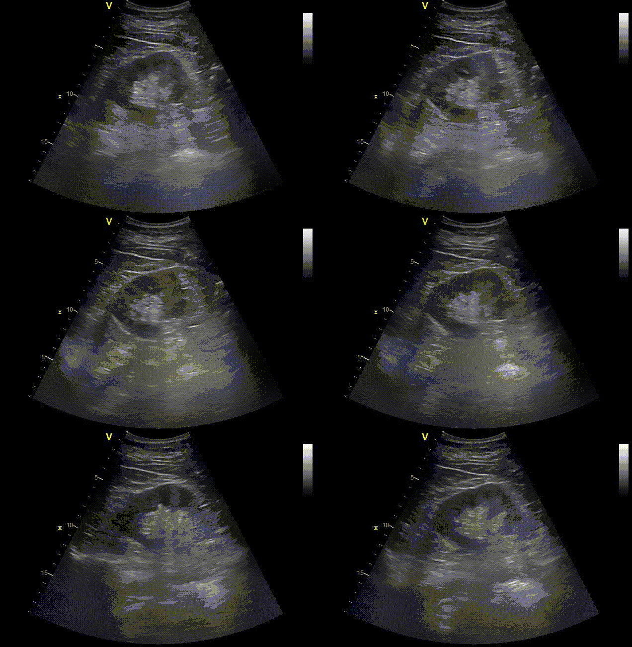
The kidneys are evaluated on abdominal ultrasound. We can see kidney stones, cysts, masses, and blocked kidneys or hydronephrosis. The kidney size can be assessed. We can look for scarring and blood flow. Some abnormalities such as masses or blocked kidneys may need further imaging with CT.
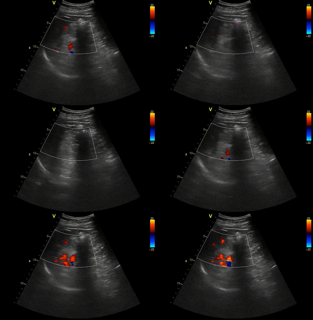
We can image the aorta. We most commonly see aneurysms of the aorta on ultrasound. The inferior vena cava is also imaged and is one of the major veins in the abdomen. We can also assess blood flow in the blood vessels with ultrasound.
Abdominal ultrasound is a comprehensive test of multiple abdominal structures. It allows us to see many abnormalities like cysts, masses, cancers, and infections to name some. Some abnormalities seen on ultrasound will require additional imaging with CT or MRI. Ultrasound is often a great start to a workup as it is relatively fast, inexpensive compared to other tests and uses no radiation.
A pelvic ultrasound can visualize several organs depending on the reason for the scan. In women, it images the uterus, ovaries, fallopian tubes, cervix, vagina, and bladder. In men, it can show the bladder, prostate, and rectum.
Doctors order pelvic ultrasounds for a wide range of reasons: investigating abnormal bleeding, pelvic pain, or irregular periods; checking for ovarian cysts, uterine fibroids, or other growths; monitoring a pregnancy in its early stages; evaluating fertility issues; looking for causes of urinary problems; or examining the prostate. It's also used to guide certain procedures, like biopsies or egg retrievals during IVF.